


The most common causes of lumbar pain are diseases of the spine, especially degenerative-dystrophic (osteochondrosis, deforming spondylosis) and overuse of the back muscles. In addition, various abdominal cavity and pelvic diseases, including tumors, can cause the same symptoms as a herniated disc and compress the root of the spine.
It is not for nothing that such patients turn not only to neurologists, but also to gynecologists, orthopedists, urologists and, above all, of course, to district or family doctors.
Etiology and pathogenesis of lumbar pain
According to modern concepts, the most common causes of loin pain are:
- pathological changes in the spine, mainly degenerative-dystrophic;
- pathological changes in the muscles, most often myofascial syndrome
- pathological changes in the abdominal organs;
- Nervous system disorders.
Risk factors for lumbar pain are:
- heavy physical activity;
- uncomfortable work posture;
- Injury;
- Cooling, drafts;
- Alcohol abuse;
- Depression and stress;
- Occupational diseases in connection with high temperatures (especially in hot shops), radiant energy, strong temperature fluctuations, vibration
Vertebral causes of loin pain include:
- Root ischemia (discogenic radicular syndrome, discogenic radiculopathy) resulting from compression of the root by a herniated disc;
- Reflex muscle syndromes, which can be caused by degenerative-dystrophic changes in the spine.
Various functional disorders of the lumbar spine can play a certain role in the occurrence of back pain, if blockages of the intervertebral joints occur due to poor posture and their mobility is restricted. Compensatory hypermobility develops in the joints above and below the block, which leads to muscle spasms.
Signs of acute compression of the spinal canal
- Numbness of the perineal region, weakness and numbness of the legs;
- Delay in urination and bowel movements;
- with compression of the spinal cord, pain relief is observed, alternating with numbness in the pelvic girdle and limbs.
Low back pain in childhood and adolescence is most commonly caused by abnormalities in spinal development. A non-overgrowth of the vertebral arches (spina bifida) occurs in 20% of adults. The examination reveals hyperpigmentation, birthmarks, multiple scars and hyperkeratosis of the skin in the lumbar area. Sometimes there is urinary incontinence, trophic disorders, weakness in the legs.
Lumbar pain can be caused by lumbarization - the transition of the S1 vertebra in relation to the lumbar spine - and sacralization - the attachment of the L5 vertebra to the sacrum. These anomalies arise due to the individual features of the development of the transverse processes of the vertebrae.
Nosological forms
Almost all patients complain of back pain, which manifests itself primarily through inflammation of the musculoskeletal system (intervertebral, rib, lumbosacral) and ligaments of the spine. Gradually an ossification develops in them, the spine loses its elasticity and functional mobility, becomes like a bamboo stick, fragile, easily injured. At the stage of pronounced clinical manifestations of the disease, the mobility of the chest when breathing, and therefore the vital capacity of the lungs, significantly decreases, which contributes to the development of a number of pulmonary diseases.
Spinal tumors
A distinction is made between benign and malignant tumors that mainly originate from the spine and metastasize. Benign spinal tumors (osteochondroma, chondroma, hemangioma) are sometimes clinically symptom-free. In the case of a hemangioma, a fracture of the spine can occur even with minor external influences (pathological fracture).
Malignant tumors, mostly metastasized, originate in the prostate, uterus, breast, lungs, adrenal gland and other organs. Pain in this case occurs much more often than with benign tumors - as a rule, persistent, painful, aggravated by the slightest movement, depriving the patient of rest and sleep. Characterized by a progressive worsening of the condition, an increase in general exhaustion, pronounced changes in the blood. X-rays, computed tomography, magnetic resonance imaging are of great importance for the diagnosis.
osteoporosis
The main cause of the disease is a decrease in the function of the endocrine glands due to an independent disease or against the background of the general aging of the body. Osteoporosis can develop in patients who take hormones, chlorpromazine, anti-tuberculosis drugs, tetracycline for a long time. Radicular diseases that accompany back pain arise from deformation of the intervertebral foramen and the spine (myelopathy) - due to compression of the radiculomedullary artery or a vertebral fracture, even after minor injuries.
Myofascial Syndrome
Myofascial syndrome is the main cause of back pain. It can occur as a result of overexertion (during vigorous physical exertion), overstretching and muscle contusions, unphysiological posture at work, reactions to emotional stress, shortening of a leg and even flat feet.
Myofascial syndrome is characterized by the presence of so-called "trigger" zones (trigger points) on which pain is caused, which often radiates to neighboring areas. In addition to myofascial pain syndrome, inflammatory muscle diseases - myositis - can also cause pain.
Often, lumbar pain occurs in diseases of the internal organs: gastric and duodenal ulcer, pancreatitis, cholecystitis, urolithiasis, etc. It can be pronounced, mimicking the picture of lumbago or discogenic lumbosacral radiculitis. However, there are also significant differences by which reflected pain can be distinguished from pain resulting from diseases of the peripheral nervous system that are due to the symptoms of the underlying disease.
Clinical symptoms for lumbar pain
Lower back pain is most common between the ages of 25 and 44. Differentiate between acute pain, which usually lasts 2-3 weeks and sometimes up to 2 months. And chronic - more than 2 months.
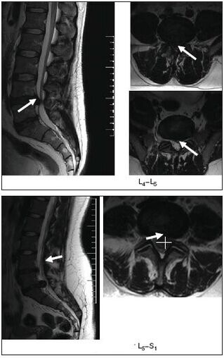
Radicular compression syndromes (discogenic radiculopathy) are characterized by a sudden onset, often after heavy lifting, sudden movements, hypothermia. Symptoms depend on the location of the lesion. At the center of the syndrome is the compression of the root by a herniated disc, which occurs as a result of dystrophic processes facilitated by static and dynamic loads, hormonal disorders, trauma (including microtraumatisation of the spine). Most often, the pathological process affects areas of the spinal roots from the dura mater to the intervertebral foramen. In addition to a herniated disc, bone growth, scarred changes in the epidural tissue and a hypertrophied ligamentum flavum can also be involved in a root trauma.
The upper lumbar roots (L1, L2, L3) rarely suffer: they make up no more than 3% of all lumbar radicular syndromes. The L4 root is affected twice as often (6%), which leads to a characteristic clinical picture: slight pain along the inner lower and anterior surface of the thigh, the medial surface of the lower leg, paresthesia (numbness, burning sensation, creeping crawling) in itArea; slight weakness of the quadriceps. Knee reflexes persist and sometimes even increase. The L5 root is most commonly affected (46%). The pain is localized in the lumbar and buttock areas, along the outside of the thigh, the antero-outer surface of the lower leg to the foot and the III-V finger. It is often accompanied by a decrease in the sensitivity of the skin of the front - outer surface of the leg and the strength in the extensor muscles of the III - V fingers. It is difficult for the patient to stand on his heel. With long-term radiculopathy, hypotrophy of the anterior tibial muscle develops and the S1 root is often affected (45%). In this case, lower back pain radiates along the outer and rear surfaces of the thigh, the outer surface of the lower leg, and the foot. The examination often reveals hypalgesia of the posterior-outer surface of the leg, a decrease in the strength of the triceps muscle and the flexor of the toe. It is difficult for such patients to stand on their toes. There is a decrease or loss of the Achilles tendon reflex.
Vertebral lumbar reflex syndrome
It can be acute and chronic. Acute Low Back Pain (LBP) (lumbago, "lumbago") occurs within minutes or hours, often suddenly due to awkward movement. A piercing, stabbing (like an electric shock) pain is localized throughout the lower back, sometimes radiating to the pelvic region andThe buttocks are enlarged when coughing or sneezing, and decrease when the patient is lying on his back, especially when the patient is comfortable. The mobility of the lumbar spine is restricted, the lumbar muscles are tense, the Lasegue symptom often occurs on both sidesPatient with legs straight on his back. At the same time, the doctor bends the affected leg at the knee and hip joints. This does not cause pain, because in this position of the leg the diseased nerve is relaxed. Then the doctor starts the leg in the hip-hip-Leaving the joint flexed, bending the knee, which tenses the sciatic nerve, causing severe pain. Acute LumbodynIt usually takes 5-6 days, sometimes less. The first attack ends faster than the following. Recurrent lumbago attacks tend to develop into chronic PB.
Atypical back pain
There are a number of clinical symptoms that are atypical for back pain caused by degenerative-dystrophic changes in the spine or myofascial syndrome. These signs include:
- the appearance of pain in childhood and adolescence;
- Back injury just before the onset of lower back pain;
- Back pain accompanied by fever or symptoms of intoxication;
- Spine;
- Rectum, vagina, both legs, belt pain;
- the association of back pain with eating, bowel movements, sexual intercourse, urination;
- necological pathology (amenorrhea, dysmenorrhea, vaginal discharge) that appeared on the background of back pain;
- increased pain in the lower back in a horizontal position and decrease in a vertical position (Razdolsky symptom, characteristic of the tumor process in the spine);
- steadily increasing pain for one to two weeks;
- Limbs and the appearance of pathological reflexes.
Survey methods
- external examination and palpation of the lumbar region, detection of scoliosis, muscle tension, pain and trigger points;
- Determination of the range of motion in the lumbar spine, areas with muscle wasting;
- Research into neurological status; Determination of tension symptoms (Lassegh, Wasserman, Neri). [Examination of Wasserman Symptom: Flexing the knee in a patient lying prone causes pain in the hip. Examination of Neri Symptom: A sharp bending of the head towards the chest of a supine patient with legs straight causes acute pain in the lower back and along the sciatic nerve.
- Examination of the state of sensitivity, reflex sphere, muscle tone, autonomic disorders (swelling, color changes, temperature and moisture of the skin);
- X-ray, computed or magnetic resonance imaging of the spine.
MRI is particularly revealing.
- Ultrasound examination of the pelvic organs;
- gynecological check;
- if necessary, additional examinations are carried out: liquor, blood and urine, sigmoidoscopy, colonoscopy, gastroscopy, etc.
treatment
Acute low back pain or exacerbation of vertebral or myofascial syndromes
Undifferentiated treatment. Soft motor mode. In the case of severe pain, bed rest for the first few days and then walking on crutches to relieve the spine. The bed should be firm, a wooden board should be placed under the mattress. A woolen scarf, an electric heating pad, bags of heated sand or salt are recommended for warming up. Ointments have a beneficial effect: Finalgon, Tiger, Capsin, Diclofenac, etc. , as well as mustard plasters, pepper plasters. Recommended UV irradiation in erythematous doses, leeches (taking into account possible contraindications), rinsing the painful area with ethyl chloride.
The anesthetic effect is achieved through electrical procedures: percutaneous electroanalgesia, sinusoidal modulated currents, diadynamic currents, electrophoresis with novocaine, etc. The use of reflexology (acupuncture, laser therapy, moxibustion) is effective; Novocaine blockade, pressure massage of trigger points.
Drug therapy includes analgesics, NSAIDs; Sedatives and / or antidepressants; Medicines that reduce muscle tension (muscle relaxants). In arterial hypotension, tizanidine should be prescribed with great caution because of its antihypertensive properties. If swelling of the spinal roots is suspected, diuretics are prescribed.
The main analgesics are NSAIDs, which are often used uncontrollably by patients when pain intensifies or recurs. It should be noted that long-term use of NSAIDs and analgesics increases the risk of complications from this type of therapy. There is currently a wide variety of NSAIDs available. For patients with pain in the spine, Diclofenac 100-150 mg / day is preferable to "non-selective" drugs in terms of availability, effectiveness and lower likelihood of side effects (gastrointestinal bleeding, dyspepsia). Indoor, intramuscular, rectal, topical, ibuprofenand ketoprofen within 200 mg, and topical and from "selective" - meloxicam within 7, 5-15 mg / day, nimesulide within 200 mg / day.
When treating NSAIDs, side effects can occur: nausea, vomiting, loss of appetite, pain in the upper abdomen. Possible ulcerogenic effect. In some cases, ulcers and bleeding in the gastrointestinal tract can occur. In addition, headache, dizziness, drowsiness, allergic reactions (rash, etc. ) are noted. Treatment is contraindicated with ulcerative processes in the gastrointestinal tract, pregnancy and lactation. To prevent and reduce dyspeptic symptoms, it is recommended to take NSAIDs during or after meals and drink milk. In addition, taking NSAIDs with increased pain in conjunction with other drugs that the patient takes to treat concomitant diseases, as in the long-term treatment of many chronic diseases, leads to a decrease in therapy compliance and, as a result, to inadequate effectiveness of the therapy.
Therefore, modern methods of conservative treatment include the mandatory use of drugs that have a chondroprotective, chondrostimulatory effect and have a better therapeutic effect than NSAIDs. The drug Teraflex-Advance, which is an alternative to NSAIDs for mild to moderate pain syndromes, fully meets these requirements. One capsule of the drug Teraflex-Advance contains 250 mg of glucosamine sulfate, 200 mg of chondroitin sulfate and 100 mg of ibuprofen. Chondroitin sulfate and glucosamine are involved in connective tissue biosynthesis and help prevent cartilage destruction and stimulate tissue regeneration. Ibuprofen has analgesic, anti-inflammatory and antipyretic properties. The mechanism of action is based on the selective blocking of cyclooxygenase (COX type 1 and type 2) - the main enzyme of arachidonic acid metabolism, which leads to a decrease in prostaglandin synthesis. The presence of NSAIDs in the Teraflex-Advance preparation helps to increase the range of motion of the joints and reduce the morning stiffness of the joints and spine. It should be noted that according to R. J. Tallarida et al. , The presence of glucosamine and ibuprofen in Teraflex-Advance offers a synergism in relation to the analgesic effect of the latter. In addition, the analgesic effect of the glucosamine / ibuprofen combination is provided by the 2. 4 times the dose of ibuprofen.
After pain relief, it makes sense to switch to Teraflex, which contains the active ingredients chondroitin and glucosamine. Teraflex is taken 1 capsule three times a day. during the first three weeks and 1 capsule 2 times / day. in the next three weeks.
The vast majority of patients show a positive trend in the form of relief from pain syndrome and a decrease in neurological symptoms when taking Teraflex. The drug is well tolerated by patients, no allergic manifestations were noted. The use of Teraflex in degenerative-dystrophic diseases of the spine is particularly useful in young patients, both in combination with NSAIDs and as monotherapy. In combination with NSAIDs, the analgesic effect occurs 2 times faster and the need for therapeutic doses of NSAIDs is gradually reduced.
In clinical practice, in lesions of the peripheral nervous system, including those associated with osteochondrosis of the spine, B vitamins with neurotropic effects are often used. Traditionally, the method of alternating administration of vitamins B1, B6 and B12, 1-2 ml, is used. intramuscularly, alternating daily. The course of treatment is 2-4 weeks. The disadvantages of this method include the use of small doses of the drug, which reduce the effectiveness of the treatment, and the need for frequent injections.
With discogenic radiculopathy, traction therapy is used: traction (even underwater) in a neurological hospital. For myofascial syndromes after local treatment (novocaine blockade, rinsing with ethyl chloride, anesthetic ointments), a hot compress is applied to the muscles for several protocols.
Chronic low back pain of vertebrogenic or myogenic origin
In the case of a herniated disc, it is recommended:
- Wearing a rigid "weight lifting belt" type corset;
- Elimination of sudden movements and inclinations, restriction of physical activity;
- physiotherapy exercises to create a muscle corset and restore muscle mobility;
- Massage;
- Novocaine blockade;
- Reflexology;
- Physiotherapy: ultrasound, laser therapy, heat therapy;
- intramuscular vitamin therapy (B1, B6, B12), multivitamins with mineral additives;
- for paroxysmal pain, carbamazepine is prescribed.
Non-drug treatments
Despite the availability of effective conservative treatments and the existence of dozens of techniques, some patients require surgical treatment.
The indications for surgical treatment are divided into relative and absolute. An absolute indication for surgical treatment is the development of the caudal syndrome, the presence of a sequestered disc herniation, a pronounced radicular pain syndrome, which, despite the ongoing treatment, does not decrease. The development of radiculomyeloid ischemia also requires urgent surgical intervention, but the indications for surgery in such cases are put into perspective, firstly by the formation of irreversible changes in the roots, and secondly, because in most cases in the course of treatment and rehabilitation measures the process is formed within approx6 months back. The same regression periods are observed for delayed operations.
Relative indications are the ineffectiveness of conservative treatment, recurrent sciatica. The duration of conservative therapy should not exceed 3 months. and lasts at least 6 weeks. It is assumed that the surgical procedure in acute radicular syndrome and ineffectiveness of conservative therapy is justified within the first 3 months. after the onset of pain to prevent chronic pathological changes in the root. A relative indication is cases of extremely pronounced pain syndrome, when the pain component changes with an increase in the neurological deficit.
For physiotherapeutic procedures, electrophoresis with the proteolytic enzyme caripazim is currently widespread.
It is known that physical therapy and massage are integral parts of the complex treatment of patients with spinal lesions. Therapeutic gymnastics aims to strengthen the body in general, increase performance, improve coordination of movements and increase fitness. At the same time, special exercises aim to restore certain motor functions.